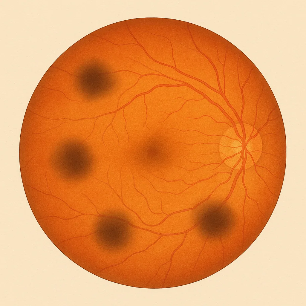

Floaters — also called myodesopsia or "eye floaters" — are small vitreous opacities perceived as dots, threads or webs that drift across the visual field. Very common, they can have very different causes: normal ageing of the vitreous, posterior vitreous detachment, high myopia, inflammation or, more rarely, haemorrhage. Dr Julien Gozlan, ophthalmic surgeon in Paris 16, reviews all these causes and the available management options.
What exactly are floaters?
The vitreous is a transparent gel that fills the eye between the lens and the retina. When small opacities — condensations of fibres, cells or pigments — form within this gel, they cast a shadow on the retina and are perceived as floaters.
These opacities can take many different forms:
- black or grey dots that move with the gaze;
- threads, rings or cobwebs (typical of posterior vitreous detachment);
- brownish or reddish spots (in the case of vitreous haemorrhage);
- a diffuse haze or cloud (inflammation, posterior uveitis).
They are particularly visible against bright backgrounds (sky, white wall, screen) and appear to "float" with a slight delay relative to eye movements.
The different causes of floaters
1. Normal ageing of the vitreous
With age, the vitreous gel naturally changes: it gradually liquefies and collagen fibres clump together into small visible condensations. This process, which begins from the age of 40–50, accounts for the vast majority of floaters in adults. It is benign and requires no treatment.
2. Posterior vitreous detachment (PVD)
The posterior vitreous detachment is the most common cause of sudden-onset floaters. After the age of 50 — earlier in myopic individuals — the membrane surrounding the vitreous separates from the retina. This phenomenon causes a sudden appearance of significant floaters, often a large floating ring (Weiss ring), and sometimes flashes of light.
In the vast majority of cases, PVD is a physiological phenomenon. However, in 10 to 15% of cases, it can lead to a retinal tear at the time of detachment — which is why any recent PVD warrants a dilated fundus examination.
3. High myopia
Individuals with high myopia (greater than –6 dioptres) have a vitreous that ages prematurely and an elongated eye whose peripheral retina is more fragile. They develop floaters earlier and are at greater risk of retinal tears and retinal detachment. Regular monitoring is essential.
4. Intraocular inflammation (uveitis)
Posterior uveitis (inflammation of the posterior segment of the eye) can cause floaters, often associated with a sensation of haziness or reduced vision. These inflammation-related floaters may be bilateral and accompanied by other systemic signs. The origin may be infectious (toxoplasmosis, herpes) or immune-mediated (sarcoidosis, Behçet's disease, etc.).
5. Vitreous haemorrhage
A vitreous haemorrhage — caused by a retinal tear, diabetic retinopathy, venous occlusion or trauma — causes a sudden appearance of reddish floaters or a dark veil over the vision. Depending on the extent of the bleeding, vision may be severely impaired. This is a situation requiring prompt management.
6. After ocular surgery or trauma
Cataract surgery, an intravitreal injection or ocular trauma can induce changes in the vitreous and the appearance of floaters. These are generally transient but warrant monitoring.
Benign or pathological floaters: how to tell the difference?
Not all floaters are equal. Some signs point towards a benign cause, while others require prompt consultation:
As a general rule, stable, few in number and long-standing floaters are benign. On the other hand, a sudden and abundant appearance in one eye — especially when associated with flashes of light, a dark veil or reduced vision — warrants prompt consultation.
To understand precisely which signs require urgent consultation, see our dedicated article: Eye floaters: when should you worry?
The ophthalmological examination
When faced with floaters, the ophthalmologist performs a dilated fundus examination. They assess the condition of the vitreous, the appearance of the peripheral retina and the possible presence of a tear or detachment. Depending on the situation:
- ocular ultrasound is used if the vitreous is opaque (haemorrhage, dense inflammation);
- vitreo-macular OCT allows detection of macular traction or an abnormal interface;
- in the case of uveitis, a targeted systemic work-up is performed according to the clinical context.
How do floaters evolve?
In the vast majority of benign cases, floaters become less bothersome over time thanks to two mechanisms:
- the opacities drift out of the central visual axis due to gravity;
- the brain gradually adapts to them (neuroadaptation).
In some patients — particularly those with high myopia or dense, central opacities — the discomfort may persist for several months or even permanently.
What treatments are available for floaters?
Monitoring and adaptation
When floaters are related to simple vitreous ageing with no associated retinal lesion, no active treatment is necessary. Fundus monitoring is recommended in the weeks following a recent PVD to ensure that no tear appears subsequently.
YAG laser vitreolysis
YAG laser vitreolysis involves vaporising vitreous opacities using a high-precision laser. It is offered in selected cases: very bothersome, accessible and central floaters in a patient with no other retinal pathology. Its efficacy is genuine in expert hands, but it is not suitable for all situations and carries some risks (transient pressure elevation, rare risk of retinal damage).
Vitrectomy
Vitrectomy is the surgical removal of the vitreous under local anaesthesia. It is rarely proposed for isolated floaters, and reserved for major, persistent symptoms, after full information about the risks: a 2022 meta-analysis (2,077 eyes) reports satisfaction in at least 90% of patients, but cataract in about one third of cases (31.7%), retinal detachment in 1.5% and endophthalmitis in 0.2%. It is, however, indicated in cases of unresolved vitreous haemorrhage or severe macular traction.
Treatment of the underlying cause
When floaters reveal an underlying cause, that cause is treated as a priority:
- a retinal tear is treated urgently by laser photocoagulation;
- uveitis is managed with anti-inflammatory or anti-infective treatment;
- a vitreous haemorrhage is monitored or operated on depending on its extent and progression.
FAQ: floaters and myodesopsia
Are floaters and myodesopsia the same thing?
"Myodesopsia" is the medical term for the perception of floaters. "Eye floaters", "spots before the eyes", "threads in the vision" — all these expressions refer to the same phenomenon of perceiving vitreous opacities.
Can floaters come back after they have diminished?
Yes. New floaters can appear with age, particularly during a PVD in the other eye or new episodes of vitreous liquefaction. Each sudden new appearance warrants an ophthalmological check-up.
Are floaters more common in myopic individuals?
Yes, significantly. Myopic eyes have a vitreous that ages prematurely and a more fragile peripheral retina. Floaters appear earlier, and the risks of PVD and retinal tear are higher. Annual monitoring is recommended for all patients with high myopia.
Is laser treatment truly effective?
In well-selected indications (central, dense, accessible opacity, at a safe distance from the retina and the lens), YAG vitreolysis yields good results. It is not universally applicable: diffuse opacities or those located too close to ocular structures cannot be treated by this technique. A personalised assessment is essential.
Can one live normally with floaters?
Yes, in the vast majority of cases. Neuroadaptation allows most patients to stop noticing them after a few weeks to a few months. In persistently very disabling cases (driving, reading, screen-based activities), a specialist discussion about therapeutic options is possible.
Do I need a referral to seek urgent consultation?
No, not necessarily. In the event of a sudden and abundant appearance of floaters, or if flashes of light or a dark veil are also present, you can go directly to the ophthalmic emergency department or call Dr Julien Gozlan's practice directly to arrange a prompt appointment.
When should you consult Dr Julien Gozlan?
Dr Julien Gozlan, ophthalmic surgeon specialising in vitreoretinal conditions, welcomes you in Paris 16 for a comprehensive assessment: dilated fundus examination, vitreo-macular OCT if required, and personalised advice on the appropriate course of action. Whether your floaters are recent or long-standing, straightforward or atypical, he will work with you to identify the cause and the most suitable treatment for your situation.
📍 Consultation at the Paris – Auteuil Ophthalmology Practice
Dr Julien Gozlan welcomes you at the Paris – Auteuil Ophthalmology Practice for the diagnosis and follow-up of floaters, posterior vitreous detachment and associated retinal conditions.
Book an Appointment on DoctolibFurther reading
- Eye floaters: when should you worry?: warning signs and emergency situations.
- Posterior vitreous detachment: the most common cause of sudden-onset floaters.
- Vitreous haemorrhage: when floaters signal bleeding.
- Retinal tear: a possible complication to look for in any PVD.