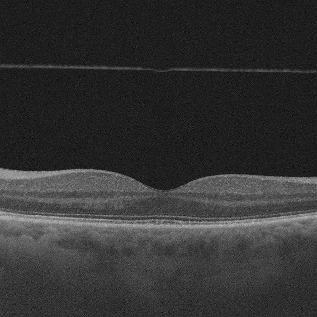

The vitreous detachment, or posterior vitreous detachment, is a very common occurrence after age 50. The transparent gel that fills the eye — the vitreous — gradually separates from the retina. In the majority of cases, the phenomenon is benign, but it can sometimes be accompanied by a retinal tear. Dr Julien Gozlan, ophthalmic surgeon in Paris 16, explains the symptoms, risks and necessary monitoring.
What are the vitreous and vitreous detachment?
Inside the eye, behind the lens, lies a transparent gel called the vitreous. This gel is surrounded by a thin membrane that adheres to the retina, especially at the periphery and at the level of the macula. With age, the vitreous liquefies and the membrane covering it gradually detaches from the retinal surface: this is posterior vitreous detachment.
This is most often a phenomenon related to the normal ageing of the eye. It can occur earlier in myopic patients, after cataract surgery or following ocular trauma.
Symptoms: when should you be concerned?
The typical signs of vitreous detachment are:
- the sudden appearance of a greater number of floaters (floating bodies);
- flashes of light at the side of the visual field, especially in the dark;
- sometimes, the perception of a ring or a moving shadow that shifts with gaze direction.
These symptoms can be alarming but do not necessarily indicate a complication. However, the appearance of a dark veil or a "curtain" progressing across the visual field is suggestive of a retinal detachment and requires emergency consultation.
Why can vitreous detachment be risky?
As it detaches, the vitreous briefly pulls on certain attachment points on the retina. In the majority of cases, this traction leaves no lasting damage. In others, it can cause a retinal tear, particularly if the retina is thin, myopic or already weakened.
A tear is a small opening in the retina through which fluid can pass and create a retinal detachment. This is why any recent vitreous detachment with clear symptoms warrants a full fundus examination.
How is the diagnosis made?
The ophthalmologist begins by measuring visual acuity and examines the fundus after pupil dilation. They look for signs of posterior vitreous detachment (floating ring, vitreous condensation) and carefully inspect the retinal periphery at the slit lamp, sometimes using a special lens.
If media transparency is poor (dense cataract, intravitreal haemorrhage), ocular ultrasound may be performed to verify that no retinal detachment is present. Macular OCT is useful when traction at the level of the macula or a complication such as a macular hole is suspected.
How long does vitreous detachment last?
This is the question almost every patient asks when they have just been given the diagnosis, and the answer often comes as a surprise: vitreous detachment is not a one-off event, but a process that unfolds over time.
The gel that fills the eye — the vitreous — separates from the retina very gradually, starting from the back of the eye. This separation occurs step by step, progressively. It is often difficult to know whether it is already complete or still partial: it is a natural process that can take several years, or even decades, to complete entirely.
This explains why no end date can be given to you. On examination, your ophthalmologist identifies a vitreous detachment in the process of forming: it is not clinically possible to confirm whether it is complete or partial.
What does change, however, is what you experience:
- flashes of light reflect the traction of the vitreous on the retina. They tend to disappear within a few days to a few weeks, once this traction ceases;
- floaters may persist for much longer. They fade as they drift out of the visual axis, and because one becomes so accustomed to them that they are no longer noticed.
A second fundus examination is performed approximately one month after the diagnosis of vitreous detachment, as the risk of retinal tear is higher in the immediate aftermath.
Typical progression and monitoring
In most cases, symptoms ease within a few weeks. The separation of the vitreous itself continues for much longer. Floaters sometimes remain visible but become less bothersome, as the brain gradually learns to ignore them. Flashes of light diminish and then disappear.
When the initial examination reveals neither a tear nor a retinal detachment, follow-up monitoring is nevertheless recommended, especially during the first few months. The patient must return for an urgent consultation if a new episode of a "shower" of floaters, repeated flashes or a veil in the visual field occurs.
What treatments are available?
Vitreous detachment itself generally requires no treatment: it is a natural stage in the life of the eye. What is treated are any potential complications:
- a retinal tear is treated urgently with laser to prevent retinal detachment;
- a confirmed retinal detachment requires surgery;
- rare cases of significant macular traction may warrant a vitrectomy.
Isolated floaters are generally not operated on. Simple measures (good hydration, visual breaks, appropriate lighting) can help in tolerating them on a daily basis.
Practical advice for patients
Following a diagnosis of vitreous detachment, it is recommended:
- to monitor for the appearance of a dark veil or a sudden drop in vision;
- to seek prompt consultation if new flashes or a shower of floaters occur;
- to continue regular ophthalmological follow-up, especially in cases of high myopia or a history of retinal detachment.
These guidelines allow for early detection of complications and timely intervention.
FAQ: frequently asked questions about vitreous detachment
Is vitreous detachment an emergency?
Vitreous detachment itself is generally not a sight-threatening emergency. However, the sudden onset of symptoms (numerous floaters, flashes of light, dark veil) should prompt prompt consultation. The aim is to verify that there is no associated retinal tear or retinal detachment, both of which require urgent treatment.
What signs should concern me in the event of vitreous detachment?
Signs that require prompt consultation are:
- the sudden shower-like onset of floaters;
- the perception of flashes of light in one eye;
- the sensation of a dark veil or curtain obscuring part of the visual field;
- an unusual or rapid drop in vision.
These symptoms may indicate a retinal tear or the onset of a retinal detachment, and must be examined without delay.
Will vitreous detachment permanently impair my vision?
In the majority of cases, vitreous detachment is a phenomenon related to the normal ageing of the eye. It mainly causes floaters and sometimes a few flashes at the outset. After a few weeks or months, the brain adapts and the disturbance diminishes considerably. Vision may remain slightly affected by certain opacities, but in the absence of a retinal complication, there is no permanent loss of central vision.
Is there a treatment to make floaters disappear?
To date, there is no simple, systematic treatment to eliminate all floaters. In the vast majority of cases, the approach relies on the brain's adaptation and the spontaneous reduction in disturbance over time.
Surgical procedures (vitrectomy) may be discussed in rare cases of major and debilitating disturbance, but they are not offered routinely, as they carry their own risks. The primary concern remains above all to verify the integrity of the retina.
Does vitreous detachment increase the risk of retinal detachment?
Yes, during the phase of recent posterior vitreous detachment, there is a risk of traction on the retina that can cause a retinal tear, which can itself lead to a retinal detachment. This risk is higher in patients with high myopia, after ocular trauma or with a history of retinal detachment in the other eye. Hence the importance of a full fundus examination at the initial stage, and sometimes a follow-up check at a later date.
Can I drive, play sport or take a flight with vitreous detachment?
In the absence of a retinal complication and if visual acuity remains adequate, driving and most activities can be continued. Floaters may, however, be more bothersome in bright light or against a light background: it is sensible to exercise extra caution while driving at first.
Vitreous detachment does not prevent air travel. However, if a tear or retinal detachment is discovered, driving, sport and sometimes travel will need to be adapted depending on the treatment required.
When should you consult Dr Julien Gozlan?
If you suddenly notice a large number of floaters, flashes of light or a veil in your visual field, it is important to seek consultation promptly. Even though vitreous detachment is often benign, only a full examination can confirm the absence of a tear or retinal detachment.
Dr Julien Gozlan, ophthalmologist in Paris 16, carries out a detailed assessment (fundus examination, OCT, and ultrasound if required) and arranges the monitoring or treatment appropriate to your situation.
📍 Consultation at the Paris – Auteuil Ophthalmology Practice
Dr Julien Gozlan welcomes you at the Paris – Auteuil Ophthalmology Practice for the diagnosis and follow-up of vitreous detachment and other retinal and vitreous conditions.
Book an AppointmentFurther reading
- Floaters: understanding "mouches volantes".
- Retinal detachment: warning signs and management.
- Macular OCT: a key examination for analysing the retina and vitreous.